Презентация "The nervous system" – проект, доклад
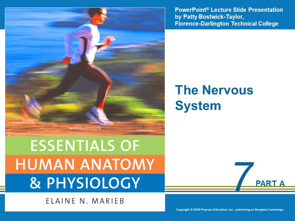 Слайд 1
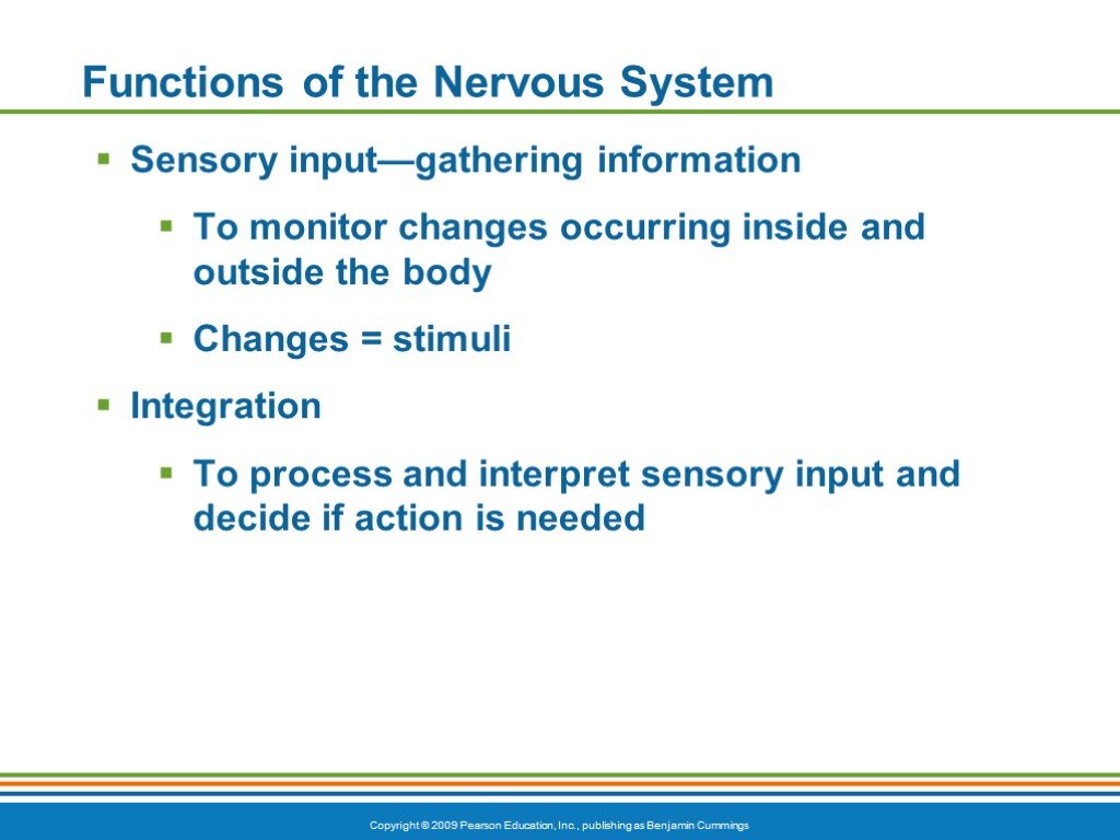 Слайд 2
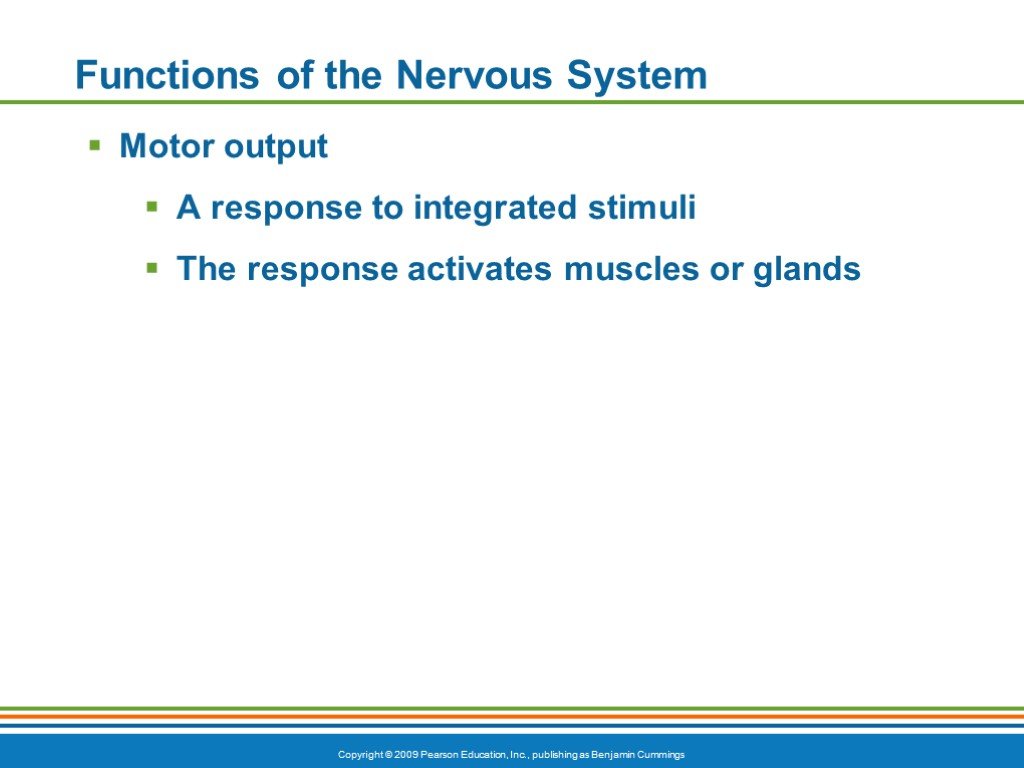 Слайд 3
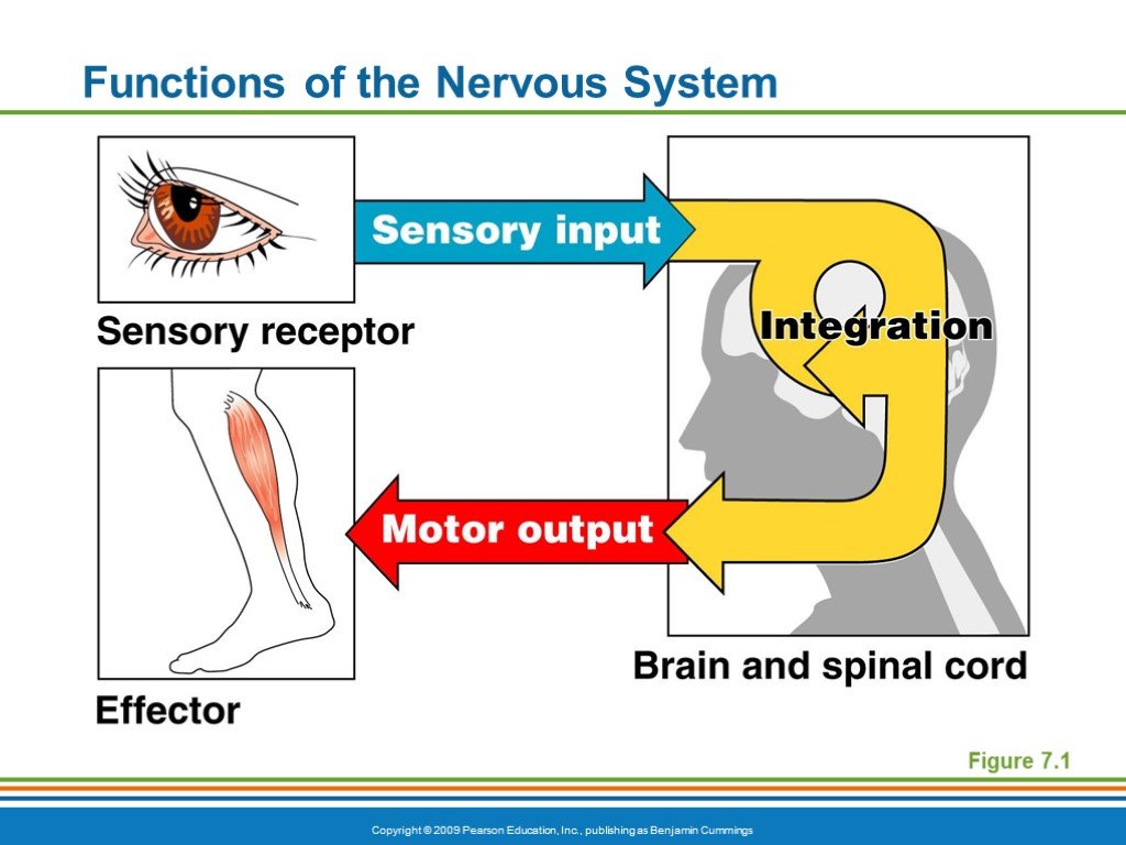 Слайд 4
 Слайд 5
 Слайд 6
 Слайд 7
 Слайд 8
 Слайд 9
 Слайд 10
 Слайд 11
 Слайд 12
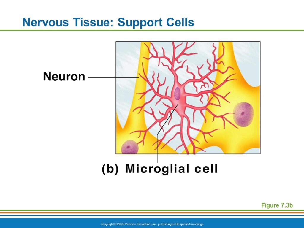 Слайд 13
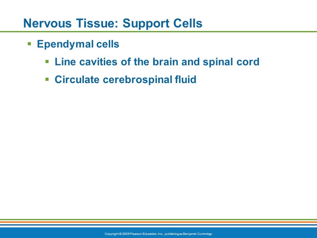 Слайд 14
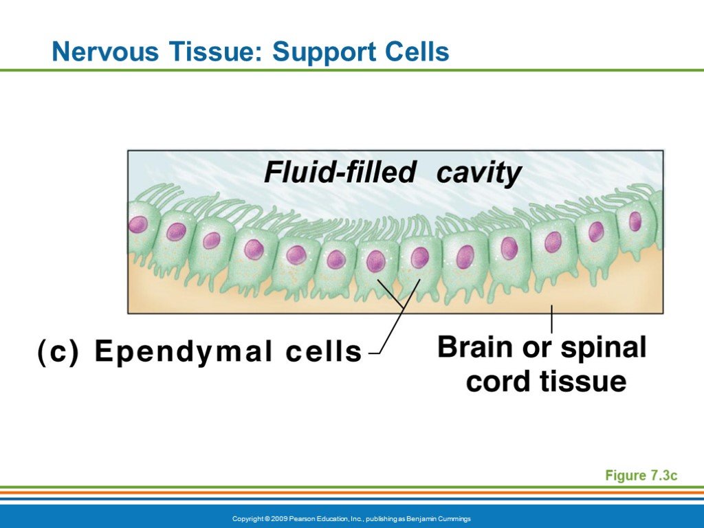 Слайд 15
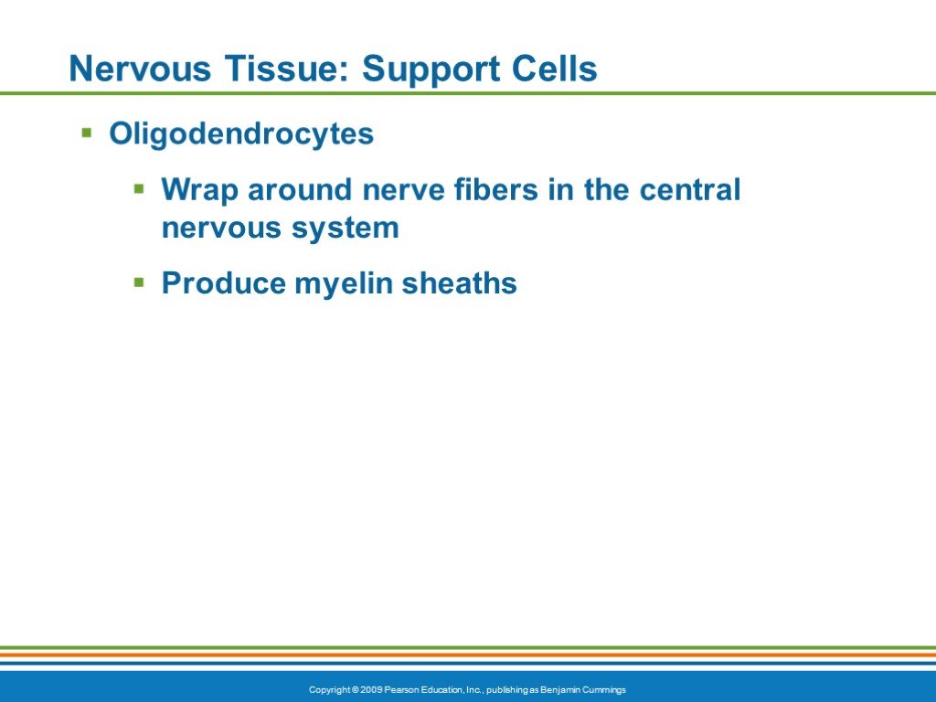 Слайд 16
 Слайд 17
 Слайд 18
 Слайд 19
 Слайд 20
 Слайд 21
 Слайд 22
 Слайд 23
 Слайд 24
 Слайд 25
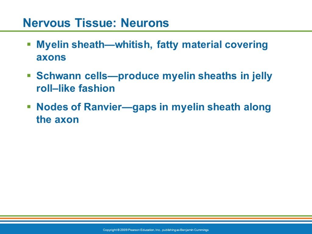 Слайд 26
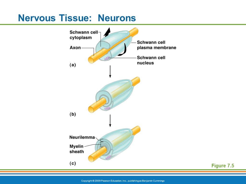 Слайд 27
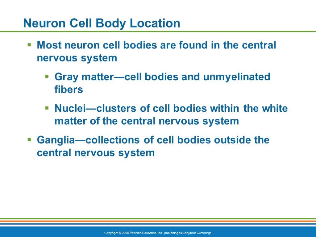 Слайд 28
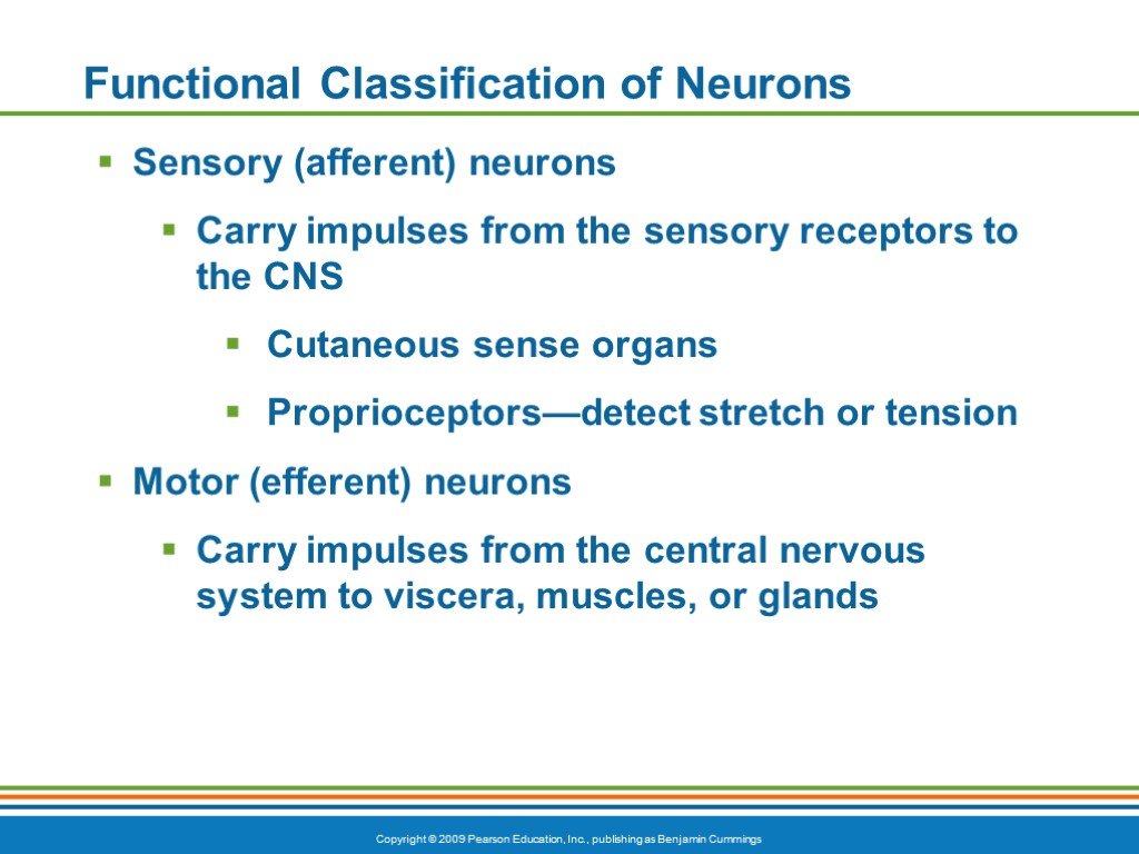 Слайд 29
 Слайд 30
 Слайд 31
 Слайд 32
 Слайд 33
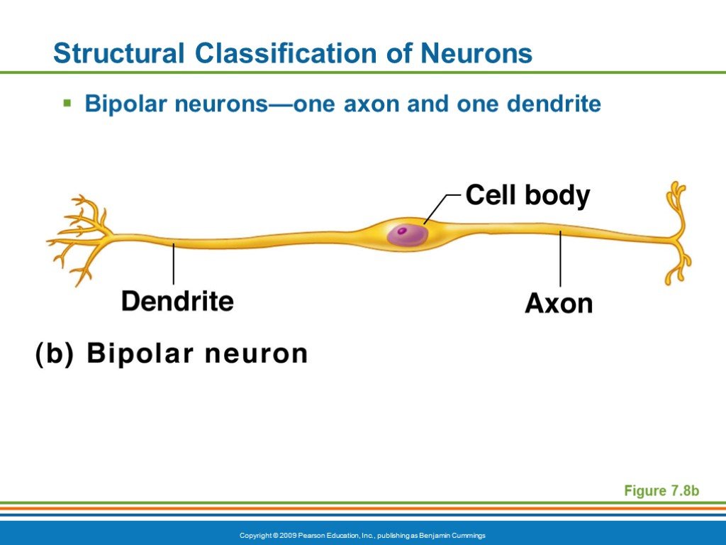 Слайд 34
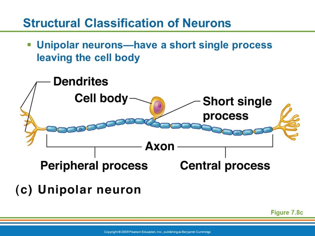 Слайд 35
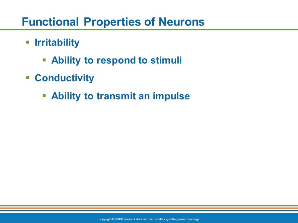 Слайд 36
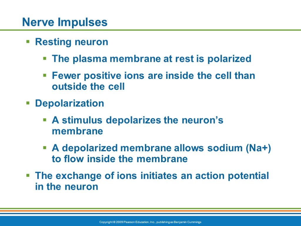 Слайд 37
 Слайд 38
 Слайд 39
 Слайд 40
 Слайд 41
 Слайд 42
 Слайд 43
 Слайд 44
 Слайд 45
 Слайд 46
 Слайд 47
 Слайд 48
 Слайд 49
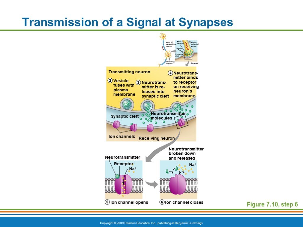 Слайд 50
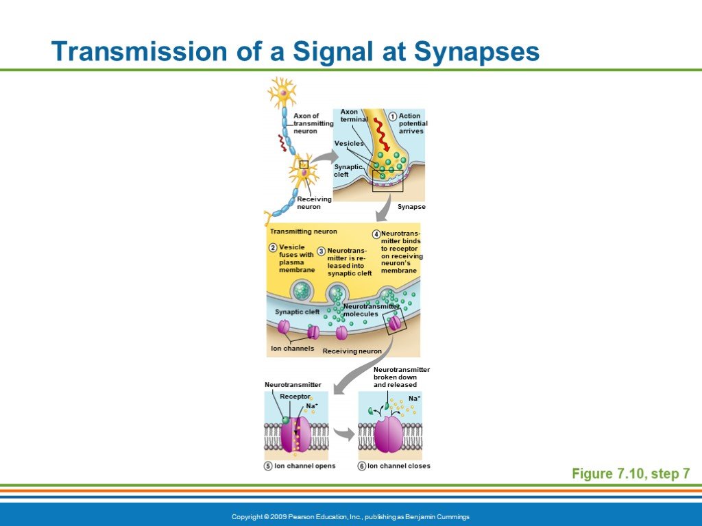 Слайд 51
Презентацию на тему "The nervous system" можно скачать абсолютно бесплатно на нашем сайте. Предмет проекта: Разные. Красочные слайды и иллюстрации помогут вам заинтересовать своих одноклассников или аудиторию. Для просмотра содержимого воспользуйтесь плеером, или если вы хотите скачать доклад - нажмите на соответствующий текст под плеером. Презентация содержит 51 слайд(ов).
Слайды презентации
Список похожих презентаций

The world work
What profession do you prefer? And Why??? Doctor? Cooker? Teacher? Architect? Signer? Firefighter? What job is the most dangerous and the most difficult??? ...
Social problems of the family
Family Problems. All families have their problems, but they may turn into a crisis if the family cannot cope or if the family does not have the resources ...
The Constitution of the United States
The Constitution was adopted on September 17, 1787, by the Constitutional Convention in Philadelphia, Pennsylvania, and ratified by conventions in ...
Interesting traditions of different peoples of the world
INDIA Festival of colors. This unusual event is taking place in New Delhi. Festival of colors - not just entertainment, it's a holiday devoted to ...Советы как сделать хороший доклад презентации или проекта
- Постарайтесь вовлечь аудиторию в рассказ, настройте взаимодействие с аудиторией с помощью наводящих вопросов, игровой части, не бойтесь пошутить и искренне улыбнуться (где это уместно).
- Старайтесь объяснять слайд своими словами, добавлять дополнительные интересные факты, не нужно просто читать информацию со слайдов, ее аудитория может прочитать и сама.
- Не нужно перегружать слайды Вашего проекта текстовыми блоками, больше иллюстраций и минимум текста позволят лучше донести информацию и привлечь внимание. На слайде должна быть только ключевая информация, остальное лучше рассказать слушателям устно.
- Текст должен быть хорошо читаемым, иначе аудитория не сможет увидеть подаваемую информацию, будет сильно отвлекаться от рассказа, пытаясь хоть что-то разобрать, или вовсе утратит весь интерес. Для этого нужно правильно подобрать шрифт, учитывая, где и как будет происходить трансляция презентации, а также правильно подобрать сочетание фона и текста.
- Важно провести репетицию Вашего доклада, продумать, как Вы поздороваетесь с аудиторией, что скажете первым, как закончите презентацию. Все приходит с опытом.
- Правильно подберите наряд, т.к. одежда докладчика также играет большую роль в восприятии его выступления.
- Старайтесь говорить уверенно, плавно и связно.
- Старайтесь получить удовольствие от выступления, тогда Вы сможете быть более непринужденным и будете меньше волноваться.
Информация о презентации
Дата добавления:24 сентября 2019
Категория:Разные
Содержит:51 слайд(ов)
Поделись с друзьями:
Скачать презентацию


